Brain microstructure mediates the relationship between Alzheimer's proteinopathies and memoryDevignes, Pruitt, Petscavage
et alAlzheimers Dement (2026) 22 (10), e71899
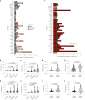
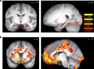
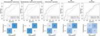
Abstract: We examined how amyloid beta (Aβ) and tau accumulation affected gray matter (GM) microstructural integrity and how such alterations impacted memory.We tested voxel-wise mediation models to examine the relationships between (1) positron emission tomography-derived Aβ and tau accumulation and GM microstructural integrity assessed with neurite orientation dispersion and density imaging, (2) GM microstructural integrity and memory controlling for Aβ and tau, and (3) the indirect effect of Aβ and tau on memory through GM microstructural integrity in 194 participants with memory impairments.Tau had a greater impact on GM microstructure than did Aβ. GM microstructural alterations in posterior cortical regions were associated with worse memory when controlling for Alzheimer's proteinopathies. GM microstructural alterations mediated the relationship between Alzheimer's proteinopathies and memory.GM microstructural alterations have functional ramifications given their mediating effect on the relationship between Alzheimer's proteinopathies and memory and their association with worse memory beyond Alzheimer's proteinopathies.Published 2026. This article is a U.S. Government work and is in the public domain in the United States. Alzheimer's & Dementia published by Wiley Periodicals LLC on behalf of Alzheimer's Association.
Age-dependent gray matter correlates of perceived financial exploitation vulnerability in cognitively unimpaired older adultsZhang, Wallman-Jones, Salminen
et alAging Brain (2026) 10, 100173
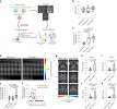
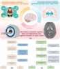
Abstract: Financial exploitation vulnerability in aging is a growing public health concern, yet its neural correlates remain poorly understood. Although psychosocial and cognitive factors confer risk, emerging evidence suggests that regional differences in brain structure may also contribute to vulnerability before overt cognitive impairment. In this study, 128 cognitively unimpaired older adults (ages 51 to 87 years, 76% female) completed structural magnetic resonance imaging and the Perceived Financial Vulnerability Scale, a self-report measure of financial exploitation risk. We examined age-stratified associations between perceived financial exploitation vulnerability and gray matter volume in relatively younger (n = 67) and older (n = 61) participants. Whole-brain voxel-based morphometry analyses revealed that higher perceived financial exploitation vulnerability was associated with lower gray matter volume in the right amygdala/anterior hippocampus (max t = 4.55, pFWE = 0.029) and left precuneus (max t = 5.40, pFWE = 0.031) among older participants, but not younger participants. In post hoc exploratory analyses modeling age as a continuous variable across the full sample, the age × perceived financial exploitation vulnerability interaction was significant for gray matter volume in the left precuneus and was observed at a trend level in the right amygdala/anterior hippocampus, with more negative associations at older ages. These cross-sectional findings identify regional gray matter correlates of perceived financial exploitation vulnerability that may vary across later adulthood.© 2026 The Authors.
Differential cross-modal functional plasticity in deaf individuals: A meta-analysis of neuroimaging studiesZuo, Li, Wang
et aliScience (2026) 29 (10), 117610
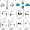
Abstract: A manifest condition of brain plasticity is cross-modal plasticity in deafness, wherein auditory-deprived cortices are recruited by intact sensory modalities. While cross-modal plasticity is well documented, the underlying principles remain debated, centering on the functional change and functional preservation principles. To quantitatively evaluate these principles, we conducted the largest coordinate-based meta-analysis to date, synthesizing 47 studies (801 deaf and 880 hearing participants) spanning linguistic and non-linguistic tasks. Deaf individuals exhibited consistent cross-modal activation in the bilateral superior temporal gyrus (STG). The left anterior STG was selectively recruited during linguistic tasks, consistent with preserved language function, whereas the left posterior and right STG were engaged across both task types, suggesting a shift toward domain-general visual processing. Cross-modal activation was lateralized, with linguistic tasks engaging the left STG and non-linguistic tasks the right. These findings support a differential cross-modal plasticity framework, wherein functional preservation and change coexist across distinct STG subregions.© 2026 The Authors.
Altered long-range visual hierarchical connectivity in internet gaming disorder: reduced V1-LOC connectivity and its association with addiction severityTian, Zhang, Wang
et alFront Psychiatry (2026) 17, 1883842
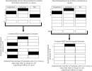
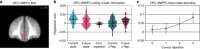
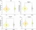
Abstract: Neuroimaging studies of internet gaming disorder (IGD) have primarily focused on reward-related and executive-control networks. However, sensory processing of addiction-related cues, particularly visual processing, may also contribute to IGD. Whether the intrinsic hierarchical organization of the visual system is altered remains unclear.The study included 35 individuals with IGD and 30 healthy controls (HCs), with an overall age range of 9-33 years. The sample was predominantly composed of adolescents. An a priori three-level visual hierarchy model comprising the primary visual cortex (V1), lingual gyrus, and lateral occipital cortex (LOC) was constructed. Functional connectivity (FC) was calculated for the V1-Lingual, Lingual-LOC, and V1-LOC pathways. A hierarchical integration index (HII), derived from V1-LOC and V1-Lingual connectivity, was calculated to assess long-range relative to local visual coupling. Group differences were assessed using MANCOVA with follow-up univariate analyses and FDR correction. Within the IGD group, associations between IAT scores and all visual hierarchical measures were examined using Pearson and covariate-adjusted partial correlations with FDR correction.V1-LOC connectivity was significantly reduced in the IGD group compared with HCs (HC: 0.364 ± 0.132; IGD: 0.130 ± 0.189; F(1,59) = 30.809; FDR-corrected p < 0.001; partial η2 = 0.343). V1-Lingual and Lingual-LOC connectivity did not show significant group differences, although their effect sizes were small-to-moderate (Cohen's d = 0.32 and 0.40, respectively). The HII was numerically lower in IGD but did not reach statistical significance (t = 1.90, p = 0.062; Cohen's d = 0.46). Within the IGD group, lower V1-LOC connectivity was associated with higher IAT scores (r = -0.479, FDR-corrected p = 0.016), and this association remained significant after controlling for age, sex, education, and mean FD (partial r = -0.447, FDR-corrected p = 0.048).IGD is associated with altered long-range hierarchical connectivity within the visual system, particularly between early visual cortex and higher-order LOC. These findings provide a sensory-level perspective complementing reward- and control-centered accounts of IGD.Copyright © 2026 Tian, Zhang, Wang, Zhang, Ma, Mei, Zhang, Lang, Wei, Han, Lv and Zhang.
Brain functional changes of suicidal ideation and behaviours: a meta-analysis of task-based fMRI studiesXu, Fang, Shao
Transl Psychiatry (2026) 16 (1)
Abstract: Suicidal ideation and behaviour are major global mental health issues and leading causes for mortality. While existing evidence indicates brain functional deficits of suicidal thoughts and behaviours, the neural underpinnings of these deficits remain unclear, possibly due to various sources of methodological heterogeneities.We conducted a pre-registered meta-analysis including 22 task-based functional magnetic resonance imaging studies that compared suicidal individuals (N = 459, 63.83% females) with non-suicidal psychiatric controls (N = 681, 52.72% females). Separate analyses were conducted in the emotion and non-emotion (cognitive and risky decision making) domains. Subgroup and meta-regression analyses addressed methodological heterogeneities including patient diagnosis, suicidal characteristics (ideation vs. behaviours) and task type.In emotion tasks, general suicidality (pooling across ideation and behaviours) and suicidal behaviours were both associated with hyperactivities in the left sensorimotor cortices and hippocampus/parahippocampus, alongside hypoactivity in the bilateral visual cortices. However, only the hypoactivity among visual areas was consistently replicated in all subgroup analyses. In non-emotion tasks, general suicidality and suicidal behaviours were both associated with hyperactivity in the left precentral gyrus and the right middle temporal gyrus, which were consistently replicated in all subgroup analyses. Meta-regression analyses further identified larger suicidality-associated functional changes in frontotemporal regions in studies with higher female proportions, and in hippocampal and temporal regions in studies with older participants.Our findings indicate that the blunted perceptual emotion processing and heightened motor system reactivity are key neural features of suicidality, which provide potential neural targets for interventions on suicidal individuals.© 2026. The Author(s).
Potential Locations for Noninvasive Brain Stimulation in Treating Poststroke Deficits: Exploration From Multimodal NeuroimagingYuan, Yang, Han
et alCNS Neurosci Ther (2026) 32 (11)
Abstract: Noninvasive brain stimulation (NIBS) shows promise for poststroke deficits (PSDs), but efficacy may be limited by inconsistent stimulation sites.By integrating neuroimaging meta-analysis, lesion-symptom mapping (LSM), and resting-state functional connectivity (FC) analysis, we employed three analytical pipelines to explore potential NIBS targets for alexia, aphasia, apraxia, ataxia, dysarthria, hemiplegia, paresthesia, and dysphagia. Pipeline 1 identified cortical regions directly associated with each PSD via coordinate-based meta-analysis using Neurosynth Compose. Pipeline 2 identified cortical regions exhibiting maximal functional connectivity with neurosynth-derived regions of interest (ROI). Pipeline 3 identified cortical regions functionally connected to ROIs from a prior large-scale LSM study. During Pipeline 2 and 3, ROIs were used for seed-to-voxel FC analysis using resting-state fMRI data from 100 healthy subjects. Identified cortical targets were mapped onto the scalp and referenced by the 10-20 system and continuous proportional coordinates.Potential cortical targets and scalp locations were identified for all eight PSDs. For instance, for hemiplegia, validated targets included primary motor and somatosensory cortices and supplementary motor area; novel targets included angular gyrus, superior frontal gyrus, inferior parietal lobule, medial prefrontal cortex, and precuneus.This multimodal approach systematically identified precise NIBS targets, confirming established sites and revealing novel regions to broaden neuromodulation strategies for poststroke recovery.© 2026 The Author(s). CNS Neuroscience & Therapeutics published by John Wiley & Sons Ltd.
Neural integration of affective prosodic and semantic cues in non-literal forms of speech understandingWittmann, Ceravolo, Mayr
et alImaging Neurosci (Camb) (2026) 4
Abstract: Emotions in speech are conveyed through both semantics and prosody-voice 'melody', yet how listeners integrate these cues in the brain remains unclear. We investigated non-literal forms of speech, such as irony and sarcasm, where understanding beyond literal meaning relies on the dynamic interplay between affective prosodic and semantic cues, alongside the theory of mind (ToM). We used functional magnetic resonance imaging while participants listened to short dialogues between two characters, which varied in prosody and semantics to convey either literal or non-literal meanings. Behaviorally, semantics and prosody interacted in shaping participants' evaluations, although the data suggest a prosody dominance effect. Non-literal speech engaged a distributed network, including the bilateral inferior frontal gyrus, temporal speech regions, and ToM areas, with ROI analyses revealing heterogeneous prosody-semantics integration profiles across regions and tasks. Taken together, our data clarify the behavioral and neural underpinnings of the integration of prosody and semantics in non-literal speech and open new avenues in this field.© 2026 The Authors. Published under a Creative Commons Attribution 4.0 International (CC BY 4.0) license.
Multi-metric resting-state fMRI reveals brain network abnormalities underlying cognitive impairment in male patients during methamphetamine abstinenceYang, Hou, Zeng
et alBMC Psychiatry (2026) 26 (1)
Abstract: Methamphetamine (MA) use disorder leads to persistent cognitive impairment, but the underlying neural mechanisms during abstinence remain unclear. This study aimed to investigate brain functional abnormalities in male methamphetamine-abstinent patients and their associations with cognitive impairment using multiple resting-state fMRI metrics.Twenty-five male MA-abstinent patients (Disease Group, DG) and twenty-nine healthy controls (Healthy Control Group, HC) were recruited, and rs-fMRI data were collected. Multimodal analysis methods, including regional homogeneity (ReHo), fractional amplitude of low-frequency fluctuations (fALFF), seed-based functional connectivity (FC), and independent component analysis (ICA), were employed to assess local spontaneous activity, hippocampal-centered functional connectivity, and large-scale resting-state network abnormalities. Cognitive function was evaluated using the Montreal Cognitive Assessment (MoCA). Group differences were compared using independent-samples t-tests, and brain-behavior relationships were analyzed using partial correlation and interaction models.Compared with the HC group, the DG group showed reduced fALFF in several regions, including the left fusiform gyrus and right inferior frontal gyrus pars triangularis, using a voxel-level threshold of p < 0.005 combined with cluster-level FWE correction at p < 0.05. Reduced ReHo was also observed in the right inferior frontal gyrus pars triangularis using a voxel-level threshold of p < 0.05 combined with cluster-level FWE correction at p < 0.05. Seed-based functional connectivity analysis revealed widespread enhanced right hippocampal functional connectivity in the DG group using a voxel-level threshold of p < 0.005 combined with cluster-level FWE correction at p < 0.05. ICA identified abnormal functional connectivity in the default mode, auditory, and somatomotor networks, with all findings surviving cluster-level FWE correction at p < 0.05. Right hippocampus-right inferior temporal gyrus connectivity was strongly positively correlated with MoCA total score in the DG group (r = 0.765, p < 0.001), and this association was significantly stronger in the DG group than in the HC group.Male MA-abstinent patients exhibit multi-layered brain functional abnormalities involving local activity, functional connectivity, and large-scale networks. These abnormalities, together with the disease-specific associations between right hippocampal functional connectivity and cognitive performance, suggest a potential role for right hippocampal networks in cognitive impairment during methamphetamine abstinence in men.© 2026. The Author(s).
Left dorsomedial prefrontal cortex hyperactivation reflects early compensation for emerging social cognition difficulties in multiple sclerosisIsernia, Pirastru, Cacciatore
et alFront Psychol (2026) 17, 1905506
Abstract: Social cognition (SC) has emerged as a relevant neurocognitive domain in people with Multiple Sclerosis (pwMS), given its impact on everyday functioning. However, the neural mechanisms supporting SC performance in pwMS remain underexplored, highlighting the need for ecologically valid assessment paradigms capable of capturing both behavioral and neural aspects of social-cognitive functioning. In this study, we addressed this gap by investigating the clinical application of an fMRI-adapted version of the Edinburgh Social Cognition Test (ESCoT) to explore the neural correlates of SC functioning in pwMS.In total, 59 subjects (27 pwMS, mean EDSS = 3.74, and 32 healthy controls) were enrolled in this study and underwent a comprehensive neuropsychological assessment, including SC tests, and the fMRI-adapted version of the ESCoT.No significant between-group differences were observed during the fMRI task performance. Across groups, the paradigm elicited activation within a network involving the bilateral middle temporal poles, left dorsomedial prefrontal cortex, and precuneus, regions consistently implicated in social-cognitive processing. Within the pwMS group, a significant association was identified between neural activation and behavioral SC performance (rho = -0.447, p = 0.032), linking poorer SC performance with greater activation of the left dorsomedial prefrontal cortex.The findings support the applicability of the ESCoT as an ecologically valid neuropsychological paradigm for capturing the neural processes underlying SC abilities in a clinical population. The observed relationship suggests that increased activation of an executive neural hub may support SC proficiency in pwMS, potentially reflecting an early compensatory strategy.Copyright © 2026 Isernia, Pirastru, Cacciatore, Rossetto, Smecca, Pedrini, Rovaris, Toselli, Blasi and Baglio.
Neuroprogression of chronic stress-induced depression via longitudinal fMRIYang, Jia, Liu
Front Psychiatry (2026) 17, 1898694
Abstract: Depression is increasingly viewed as a progressive neurobiological trajectory, yet the spatiotemporal transition from early mood dysregulation to chronic network failure, and its relation to disease-related functional decline, remains unclear. Using a chronic unpredictable mild stress (CUMS) mouse model, we aimed to map the longitudinal brain transformation from local spontaneous activity to global network dysfunction via multi-parametric MRI.Longitudinal functional magnetic resonance imaging (fMRI) was performed on a cohort of CUMS mice (n = 7) across three longitudinal time points: Day 0 (baseline), Day 5 CUMS, and Day 10 CUMS. We integrated four analytical approaches: fALFF, static functional connectivity, graph theory, and dFC/dALFF.Depression progression followed a hierarchical spatiotemporal dissolution. fALFF revealed sequential breakdown of local hubs in caudoputamen, medial prefrontal cortex, and hippocampus, suggesting involvement of motor-motivational and executive deficits. sFC showed sensory-limbic uncoupling between caudoputamen and visual areas, and altered mPFC-AON connectivity and hippocampal prefrontal (HPF-PL) dysconnectivity, indicating limbic-olfactory loop disruption. Graph theory indicated preserved global node efficiency but widespread loss of node local efficiency and clustering. Dynamic analyses uncovered a shift from flexible network reconfiguration to dynamic rigidity.Longitudinal CUMS exposure was associated with progressive alterations in functional network organization. The prefrontal olfactory dysconnectivity and onset of dynamic rigidity represent candidate multi-scale and longitudinal imaging features that warrant future validation.Copyright © 2026 Yang, Jia and Liu.
Differences in Near-Infrared Light Spectral Characteristics and Cranial MRI Parameters in Patients with Major Depressive DisorderHuang, Wan
Psychiatry Clin Psychopharmacol (2026) 36 (2), 143-149
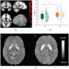
Abstract: This study aimed to evaluate differences in cerebral hemodynamics and regional brain activity using functional near-infrared spectroscopy (fNIRS) and resting-state functional MRI in patients with major depressive disorder compared with healthy controls.Sixty patients with major depressive disorder admitted to our hospital from June 2022 to June 2023 were selected as the major depressive disorder (MDD) group, and another 60 healthy controls with physical examination in our hospital during the same period were selected as the healthy control group. The subjects were subjected to functional infrared spectroscopy imaging, and the integration values and center of gravity parameters in the frontal area and bilateral temporal areas were compared between the 2 groups of subjects. MRI scans were performed on all subjects to obtain brain perfusion images.The integral values of the prefrontal region and bilateral temporal regions in the MDD group were reduced, and the center of gravity value of the prefrontal region was raised in the MDD group. Regional homogeneity values in the left temporal lobe, frontal lobe, cingulate gyrus, and left precentral gyrus decreased in patients with MDD. Regional homogeneity values increased in the right frontal lobe, anterior cingulate gyrus, right thalamus, and left insular lobe. Multiple Logistic Regression Analysis showed that years of education, forehead score value in functional near-infrared spectroscopy index, ReHo values in the right thalamus and right anterior cingulate gyrus were risk factors for patients with MDD, and ReHo values in the left precentral gyrus, left frontal lobe, and cingulate gyrus were protective factors for patients with MDD (P < .05).Near-infrared light spectroscopy imaging parameters and cranial MRI cerebral blood flow differ in patients with MDD.2026 Jianjie Huang, Dandan Wan.
Gray matter abnormalities associated with long-term opium use: A voxel-based morphometry studyEbrahimi, Pourrahimi, Amiri
Drug Alcohol Depend Rep (2026) 21, 100485
Abstract: Chronic raw opium use can result in opioid use disorder (OUD). Although voxel-based morphometry (VBM) studies have demonstrated decreased gray matter volume (GMV) in heroin-induced OUD, the structural impacts of long-term raw opium consumption remain poorly understood. This study investigated GMV alterations in men with opium-induced OUD and their association with use duration.High-resolution T1-weighted MRI scans were acquired from 28 men with opium-induced OUD (mean use duration 17.89 ± 5.85 years) receiving opium tincture maintenance and 25 age- and education-matched healthy male controls. Whole-brain VBM (CAT12/SPM12) was controlled for age, total intracranial volume, and nicotine use. Analysis used a voxel-level uncorrected threshold of p < 0.05 with cluster-level FDR correction (p < 0.05), along with stricter thresholds (uncorrected p < 0.001, TFCE with FWE), and small-volume correction (SVC). Partial correlation and multiple linear regression investigated clinical associations.Whole-brain analysis identified a left-hemisphere cluster of reduced GMV (cluster-level FDR-corrected p = 0.009). No clusters survived p < 0.001 or TFCE with FWE correction. Reductions in the left insula, inferior frontal gyrus, and parahippocampal gyrus remained significant after SVC. Left insula GMV was negatively associated with duration of opium use (partial r = -0.45, p = 0.027; β = -0.48, p = 0.027 in regression).Prolonged raw opium use is associated with decreased GMV in areas involved in craving and impulse control. Given that the whole-brain findings were not confirmed by more stringent statistical approaches, the results should be considered exploratory and necessitate replication in larger samples.© 2026 The Author(s).













.svg "Stanford University")